Figure 1.
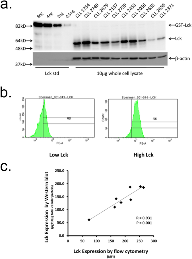
Variable Lck expression levels in CLL cells determined by Western blot and flow cytometry. (a) Western blot analysis of Lck expression in lysates of purified CLL cells. 10 μg of protein derived from lysates of CD19-purified CLL cells was separated by SDS-PAGE and immunoblots probed with either anti-Lck mAbs or anti-β-actin (as loading control). Varying amounts of purified recombinant Lck protein were used as standards to determine Lck levels in cell lysates. (b) Flow cytometry histograms of Lck expression in CLL cells derived from cases with known high and low levels. (c) Correlation of Lck expression in CLL cells determined by Western blot versus flow cytometry.

