Figure 2.
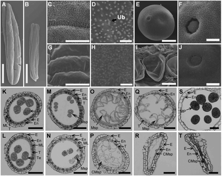
Defective development of the ms6021 anther surface and pollen wall. (A,B) Anthers of wild-type (A) and ms6021 (B) at the mature pollen stage. (C,G) SEM analysis of the anther surface of wild-type (C) and ms6021 (G) at the mature pollen stage. (D,H) SEM analysis of the inner surface of wild-type (D) and ms6021 (H) at the mature pollen stage. (E,F,I,J) SEM analysis of the pollen grain (E,I) and pollen surface (F,J) of wild type (E,F) and (I,J) at the mature pollen stage. (K) to (T) Cytological comparison of anther development in wild type and ms6021 at different stages. The anthers of wild type are shown in (K,M,O,Q and S); and of the ms6021 mutant are shown in (L,N,P,R and T) tetrad stage (K,L); uninucleate stage (M,N); large vacuole stage (O,P); binucleate stage (Q,R); mature pollen stage (S,T). CMsp, collapsed microspore; E, epidermis; En, endothecium; ML, middle layer; MP, mature pollen; Msp, microspore; T, tapetum; Te, tetrad; Ub, ubisch body. Bars = 1 mm in (A,B), 20 µm in (C) to (E) and (G) to (I), 5 µm in (F,J), and 50 µm in (K) to (T).

