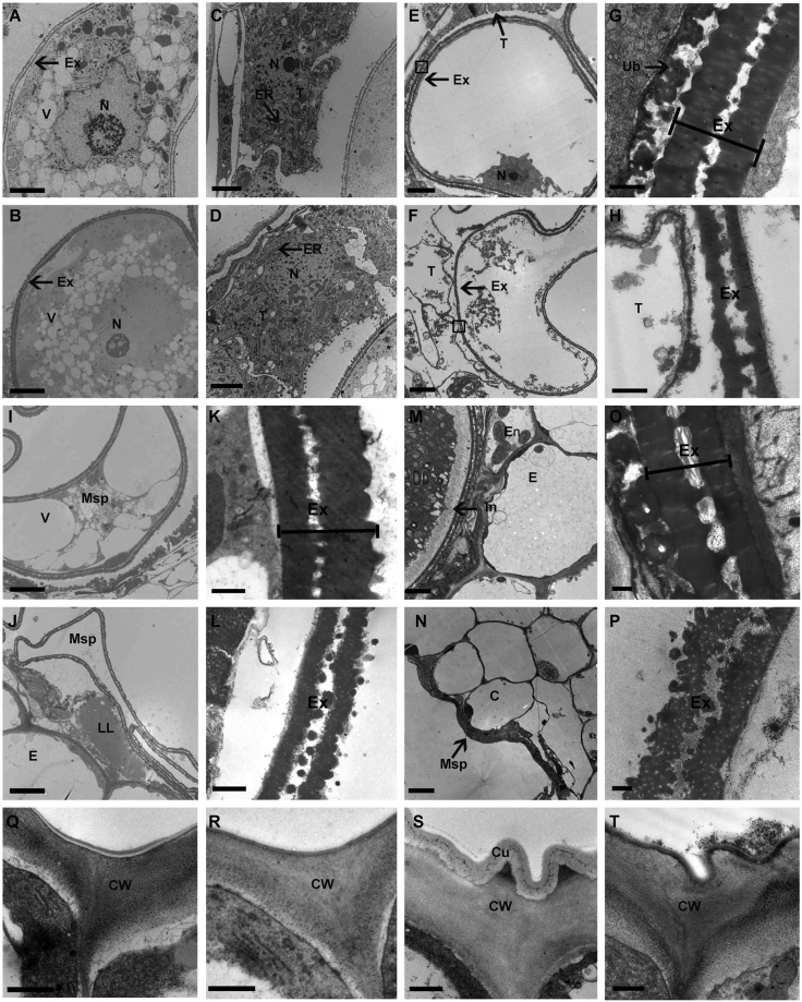
Figure 3.
TEM analysis of anthers from wild-type and ms6021. (A) to (D) Microspore (A,B) and tapetum (C,D) of anthers from wild type (A,C) and the mutant (B,D) during the uninucleate stage. (E) to (H) Microspore (E,F) and pollen exine (G,H) of anther from wild-type (E,G) and mutant (F,H) at large vacuole stage. (I) to (L) Microspore (I,J) and pollen exine (K,L) of anthers from wild type (I,K) and the mutant (J,L) at the binucleate stage. (M) to (P) Anther wall (M,N) and pollen exine (O,P) of anthers from wild type (M,O) and the mutant (N,P) at the mature pollen stage. (Q) to (T) Anther epidermal surface of wild type (Q,S) and the ms6021 mutant (R,T) at the uninucleate stage (Q,R) and mature pollen stage (S,T). C, cavity for dehiscence. Cu, cuticle; CW, cell wall; ER, endoplasmic reticulum; Ex, exine; In, intine; LL, lipidosome-like; Msp, microspore; N, nucleus; T tapetum; Ub, ubisch body; V, vacuole. G and H were zoomed form black solid boxes region in E and F respectively. Bars = 5 µm in (A,B,E,F,I,J,M and N), 2 µm in (C,D), and 0.5 µm in (G,H,K,L,O and P) and (Q) to (T).

