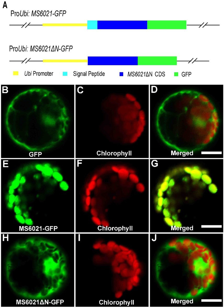
Figure 7.
Subcellular localization analysis of MS6021. (A) Diagram of the full-length constructs of MS6021 cDNA and signal region deleted cDNA fused to GFP under the control of the maize ubiquitin promoter. (B) to (D) A maize protoplast expressing empty pJIT163-GFP showing green fluorescence (B), chlorophyll autofluorescence (C), and the merged signals (D) of (B) and (C). (E) to (G) A maize protoplast expressing fused MS6021-GFP showing green fluorescence (E), chlorophyll autofluorescence (F), and the merged signals (G) of (B) and (C). (H) to (J) A maize protoplast expressing empty fused MS6021ΔN-GFP showing green fluorescence (H), chlorophyll autofluorescence (I), and the merged signals (J) of (H) and (I). Bars = 10 µm.

