Figure 1.
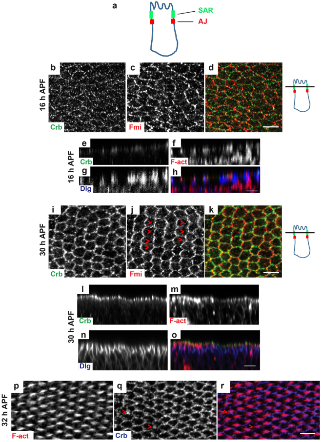
Crb displays a dynamic redistribution during pupal wing development. (a) Schematic drawing of a Drosophila epithelial cell, showing the position of the subapical region (SAR, in green) and of the adherens junctions (AJ, in red). (b–d and i,k) Crb (green) and Fmi (red) distribution in pupal wings at 25 °C at 16 h (b–d) or 30 h (i,k) APF; Red arrowheads in panel J show the Fmi zig-zag pattern oriented orthogonally to the PD axis. (e–h and l–o) Orthogonal sections of pupal wings at 16 h (e–h) or 30 h APF (l–o) stained for Crb (green), F-actin (red) and Dlg (blue). (p–r) Pupal wing at 32–34 h APF stained for Crb (blue) and F-actin (red). Red arrowheads in panel Q show Crb accumulation at the bottom of emerging hair. On the right of panels B–D and I–K drawn orthogonal views of a wing epithelial cell where the focal plane positions of the confocal image projections in the left panels are indicated (black line). All images are maximal projections of 2 up to 6 optical sections (every 0.2 μm). Distal is right, proximal left. Scale bar: 10 μm.

