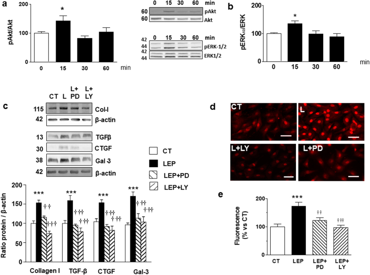
Figure 2.
Effect of leptin on Akt and MEK pathways and impact of inhibition of Akt and MEK pathways on profibrotic protein factors and ROS levels in cardiac myofibroblasts. Protein levels of (a) pAkt/Akt and (b) pERK 1/2/ERK1/2 stimulated by leptin (100 ng/mL) for indicated time intervals. Cardiac myofibroblasts stimulated for 24 hours with leptin (100 ng/mL) in the presence or absence of the inhibitors of either MEK (PD98059; PD; 25 × 10−6 mol/L) or Akt (LY294002; LY; 20 × 10−6 mol/L) pathways for 24 hours. (c) Protein levels of collagen type I, TGF-β, CTGF and galectin-3. (d) Representative microphotographs in cells labeled with DHE and (e) Quantification of total superoxide anions and. Bar graphs represent the mean ± SD of 3–4 assays in arbitrary units normalized to β-actin. *p < 0.05; ***p < 0.001 vs. control. ††p < 0.01; †††p < 0.001 vs. leptin.

