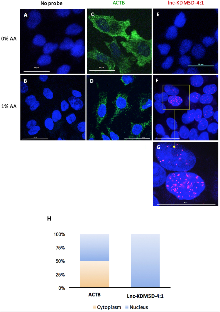
Figure 4.
Subcellular localization of lnc-KDM5D-4:1 in HepG2 cells. (A,B) Cells without probes (negative control). (C,D) actin beta (ACTB fluorescence in situ hybridization (ACTB-FISH) (green) used as housekeeping gene and positive control for the RNA FISH assay. (F,G, and Supplementary Video S1) RNA FISH shows the nuclear localization of lnc-KDM5D-4:1 (red). Each probe was tested with (B,D,F,G, and Supplementary Video S1) or without (A,C,E) addition of 1% of acetic acid (AA) at the cell fixation step. This resulted in no observable nuclear signal for lnc-KDM5D-4:1 when AA is not added to the fixation solution (E). (H) Real-time PCR lnc-KDM5D-4:1 transcript expression results from nuclear and cytoplasmic RNAs isolated separately from HepG2 cells before reverse transcription, then real-time PCR. Results show a nuclear expression, exclusively, of lnc-KDM5D-4:1 in comparison to ACTB which is approximately half nuclear and half cytoplasmic. Microscopy confocal images represent three independent experiments (n = 3). Confocal microscope magnification: 60x.

