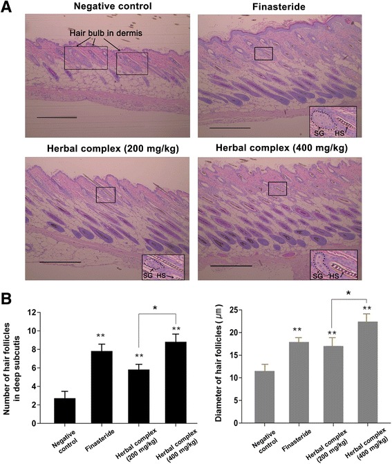
Fig. 3.

Histologic analysis of hair follicle growth in C57BL/6 mice. Skin samples were fixed in 10% paraformaldehyde, embedded in paraffin, sectioned, and stained with hematoxylin and eosin. a Hairs in the negative control group on day 25 were in the early anagen phase (hair bulb in the dermis). In contrast, the hair follicles in the experimental groups and in the finasteride group were at least in the anagen IIIc-IV phase, showing maximal size of hair bulb, hair follicle deep in the subcutis, and newly formed hair shaft reaching the level just below the sebaceous gland. The higher dose (400 mg/kg) used in the experimental group resulted in deeper hair bulb and larger hair follicle than observed in the low dose (200 mg/kg) group. (×100 objective magnification, scale bar = 100 μm). b The hair follicle counts in deep subcutis and the diameter of hair follicles. Data are presented as the mean ± SD. **p < 0.001 compared with negative control, *p < 0.05 herbal complex 200 mg/kg vs. 400 mg/kg group. Herbal complex = Houttuynia cordata Thunb, Perilla frutescens Britton var. acuta, and Green tea. SG, sebaceous gland; HS, hair shaft
