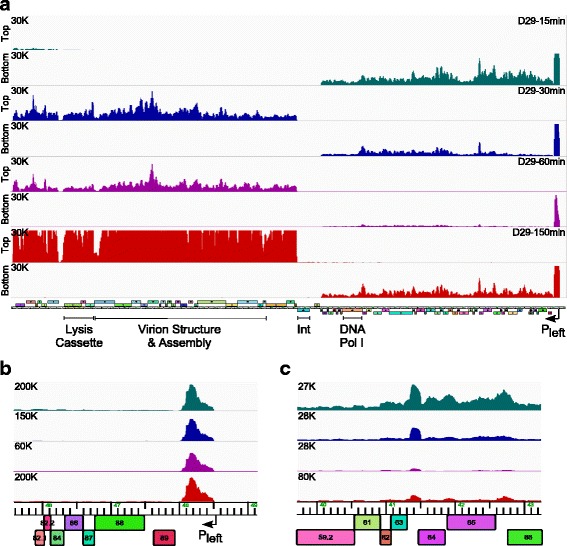
Fig. 4.

Transcriptomic analysis of D29. a. Strand specific RNAseq analysis of D29 (MOI = 3) infected M. smegmatis mc2155. Time points after adsorption are indicated on the upper right; 15 min (teal), 30 min (blue), 60 min (purple), 150 min (red). At the left are scale maxima and indication of top or bottom strand. The D29 map is shown at the bottom. b. Detailed view of the RNAseq reads from the bottom strand at the extreme right end of the D29 genome. Samples are color coordinated with panel A, but note the scale difference from panel a. c. A detailed view of the RNAseq reads (bottom strand) of the transcribed intergenic region between 63 and 64
