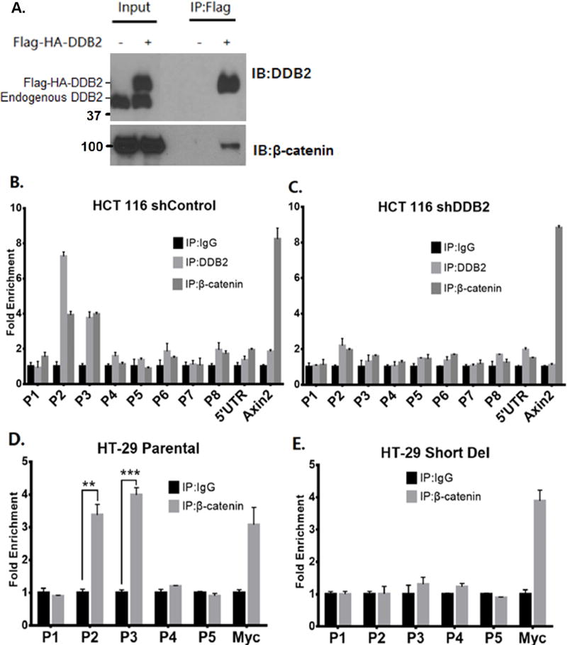
Figure 3. DDB2 recruits β-catenin onto Rnf43 upstream regulatory region.
(A) Nuclear extracts of HCT 116 cells expressing empty vector or Flag-HA-DDB2 were immunoprecipitated by ANTI-FLAG M2 affinity gel and analyzed by Western Blotting using DDB2 and total β-catenin antibodies. IP: Immunoprecipitation; IB: Immunoblotting. (B and C) ChIP assay was performed to analyze the local enrichment of DDB2 and β-catenin across the upstream regulatory region and part of 5’ UTR of Rnf43gene in HCT 116 cells expressing either shControl (B) or shDDB2 (C). The relative fold enrichment was quantified by normalization to input. And the fold enrichment of IgG is set as 1 for all primer sets. A primer set which amplifies the TCF/ β-catenin binding site on Axin2 intron1 (Cell Signaling) is used as positive control for β-catenin immunoprecipitation. (D and E) ChIP assay was performed to analyze the local enrichment of β-catenin from P1 to P5 regions of Rnf43gene in HT-29 Parental (D) and Short Del (E) cells. The relative fold enrichment was quantified by normalization to input. And the fold enrichment of IgG is set as 1 for all primer sets. A primer set which amplifies the TCF/ β-catenin binding site on c-Myc promoter is used as positive control. N = 3, Error bars indicate SD. **: P <0.01. ***: P <0.001

