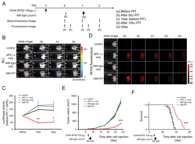
Figure 5. In vivo effect of NIR-PIT for MOC2-luc tumor in a unilateral tumor model.
(A) NIR-PIT regimen. Bioluminescence and fluorescence images were obtained at each time point as indicated. (B) In vivo BLI of tumor bearing mice in response to NIR-PIT. Before NIR-PIT, tumors were approximately the same size and exhibited similar bioluminescence. The tumor treated by NIR-PIT showed decreasing luciferase activity after NIR-PIT. (C) Quantitative luciferase activity showed a significant decrease in NIR-PIT tumors (n ≧ 8, *p < 0.05 vs. other groups, **p < 0.01 vs. other groups, by Tukey’s t test with ANOVA). (D) In vivo IR700 fluorescence real-time imaging of tumor-bearing mice in response to NIR-PIT. The tumor treated by NIR-PIT showed decreasing IR700 fluorescence immediately after NIR-PIT. (E) Tumor growth was significantly inhibited in the NIR-PIT treatment groups (n ≧ 8, ***p < 0.001 vs. other groups, Tukey’s test with ANOVA). (F) Significantly prolonged survival was observed in the NIR-PIT treatment group (n ≧ 8, ***p < 0.001 vs. other groups, by Log-rank test).

