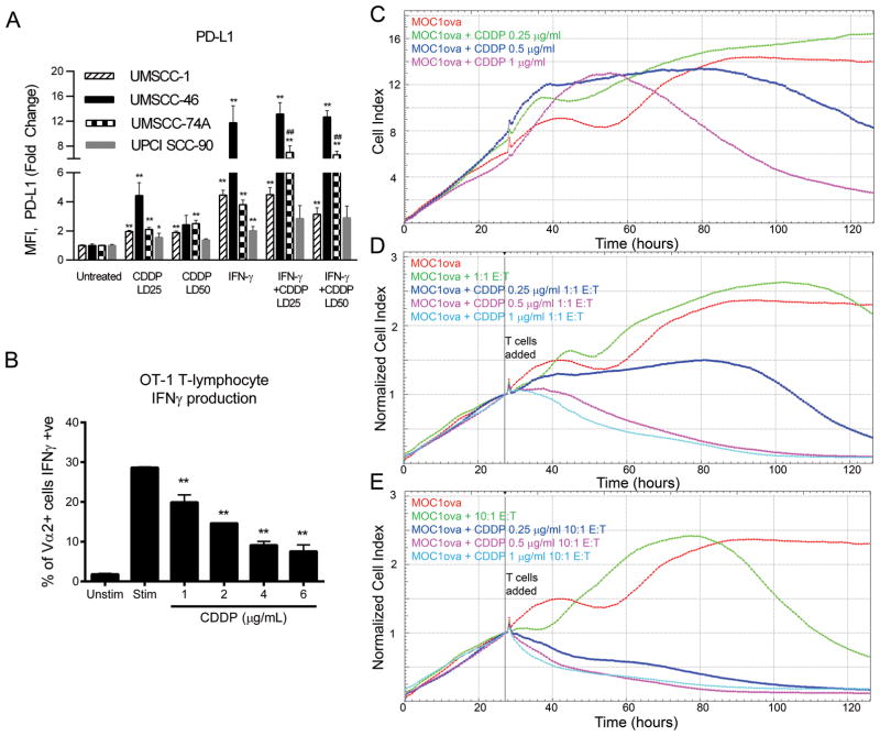
Figure 2. Cisplatin upregulates PD-L1 on tumor cells, impairs T-cell IFNγ production in a dose-dependent manner, and enhances antigen-specific T-cell killing.
A, HNSCC cells were treated with cisplatin and/or IFNγ and cell surface PD-L1 was measured by flow cytometry. Cell lines: UMSCC-1 (striped), UMSCC-46 (solid black), UMSCC-74A (bricked), and UPCI SCC90 (solid gray). Data are mean + SEM from at least two independent experiments performed in triplicate, * p <0.05, ** p <0.01 vs. untreated, ## p <0.01 vs. IFNγ or CDDP alone, by one-way ANOVA with post-hoc Tukey’s multiple comparison test. B, OT-1 T cells were cultured with or without varying concentrations of cisplatin, then left unstimulated or pulsed with SIINFEKL peptide. A flow cytometry-based IFNγ capture assay was then used to measure production of IFNγ by antigen-specific T cells. Data are mean + SEM from one triplicate experiment, ** p <0.01 vs. stimulated without prior cisplatin by one-way ANOVA with post-hoc Tukey’s multiple comparison test. C–E, Ovalbumin-expressing MOC1 tumor cells (MOC1ova) were allowed to grow in 96-well plates in the presence or absence of cisplatin for 24 hours prior to adding antigen-specific OT-1 T cells (T: target MOC1ova cells and E: effector OT-1 T cells). Impedance measurements were taken over time to determine tumor cell viability. Graphs are representative of two independent experiments done in triplicate. Different doses of CDDP are indicated by color. D and E, Impedance lines are graphed as averages of 3 replicates that have been normalized to a cell index of 1.0 at 26 hours when CTLs were added. CTLs were exposed to cisplatin along with MOC1ova cells starting at 26 hours but were not pretreated with cisplatin.

