
Fig. 4.
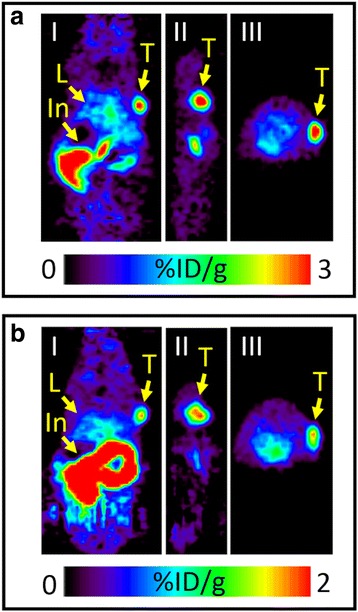
PET images of pretargeted cetuximab in A431 tumour-bearing mouse in group A (a) and pretargeted trastuzumab in BT-474 tumour-bearing mouse in group A (b). a 75 μg of TCO-cetuximab (3.1 nmol of TCO) was administered 72 h prior to the injection of [18F]TAF (4.3 ± 0.1 nmol) intravenously (n = 4), and b 20 μg of TCO-trastuzumab (0.65 nmol of TCO) was administered 72 h prior to the injection of [18F]TAF (1.4 ± 0.1 nmol) intravenously (n = 4). Coronal (I), sagittal (II), and transverse (III) planar images intersect the centre of the tumours. Arrows indicate the locations of the tumour (T), liver (L), and intestines (In)