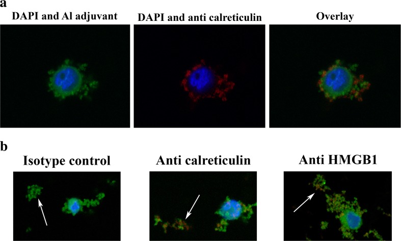
Fig. 5.
a Confocal images of THP-1 cells co-cultured with lumogallion-labelled Adju-Phos and stained with anti-calreticulin. THP-1 cells were co-cultured over night with 200 μg/ml lumogallion-labelled AdjuPhos. The next day, the cells were stained with APC-labelled anti-calreticulin, washed with PBS, fixed in PFA and mounted using ProLong® Gold Antifade Reagent. A confocal z-stack was made and the centre of a cell is shown in the figure. To the left: nuclear (blue, DAPI) and Adju-Phos staining (green, lumogallion). In the middle: nuclear (blue, DAPI) and anti-calreticulin staining (red, APC). To the right: an overlay image showing nucleus (blue), Adju-Phos adjuvant (green) and anti-calreticulin (red) staining. b Confocal images of aggregates of lumogallion-labelled Adju-Phos stained with anti-HMGB1 and anti-calreticulin. Cells were co-cultured with lumogallion-labelled Adju-Phos as described in Fig. 3a. After co-culturing, the cells were stained with APC-labelled antibodies against calreticulin, HMGB1 or an isotype control antibody; washed with PBS; fixed in PFA and mounted using ProLong® Gold Antifade Reagent. Confocal z-stacks were made, and a section slightly below the cell centre showing a cell and non-endocytosed Adju-Phos aggregates is shown in the figure. Arrows pointing at non-endocytosed Adju-Phos aggregates. To the left: an overlay image after staining with the isotype control antibody. In the middle: an overlay image after staining with anti-calreticulin. To the right: an overlay image after staining with anti-HMGB1. Nucleus stained with DAPI (blue), Adju-Phos adjuvant stained with lumogallion (green) and antibodies labelled with APC (red)

