Figure 5.

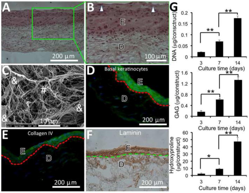
Formation of skin substitutes containing epidermal (E) and dermal (D) compartments after 2-week culture of assembled 3D cell/nanofiber constructs, in which fibroblast seeding and nanofiber electrospinning were alternated for 10 layers followed by 5 layers of keratinocyte and electrospinning alternation. (A, B) Optical microscopic image of H & E stained cross-sections of skin substitutes with the presence of granular cells (arrow). (C) SEM micrograph of cross-sections of the dermal compartment of skin substitutes composed of ECM fibers (#) and fibroblasts (&). (D) Fluorescent image of cross-sections of skin substitutes immunofluorescently stained for basal keratinocytes (green) and nuclei (blue). (E) Fluorescent image of cross-sections of skin substitutes immunofluorescently stained for type IV collagen (green) and nuclei (blue). (F) Optical image of the cross-sections of skin substitutes immunohistochemically stained for laminin (brown). Broken lines in (D-F) indicate the separation between epidermis and dermis. (G) Quantitative analyses of cell proliferation and new ECM deposition in skin substitutes (n=4). * p <0.05. ** p <0.001
