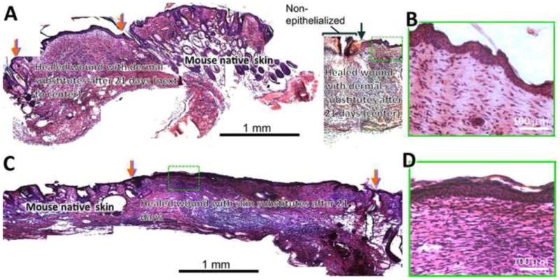
Figure 8.

Histology of the wounds treated with dermal (A, B) and skin (C, D) substitutes after 3 weeks. The wounds grafted with dermal substitutes fabricated from 15 alternating layers of fibroblasts and nanofibers did not completely close by 3 weeks with non-epithelialized center (A, right) and pronounced contraction (A, left). Red arrows indicate the wound edge with substitutes and black arrow indicates the edge of re-epithelialization. Close examination (B) revealed the regeneration of epidermal structure on top of dermal substitutes upon epidermis migration from wound edge. Wounds grafted with skin substitutes (10 alternating layers of fibroblasts and nanofibers followed by 5 alternating layers of keratinocytes and nanofibers) completely closed (C) with a full coverage by stratified epidermis (D). Red arrows indicate the wound edge with substitutes.
