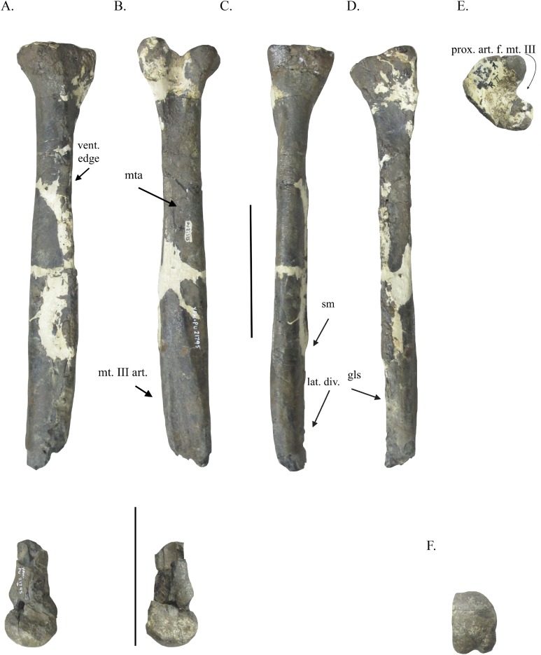
Figure 2. Metatarsal IV of YPM VPPU.021795.
Metatarsal IV in lateral (A), medial (B), dorsal (C), ventral (D), proximal (E), and distal (D) views. Abbreviations: vent. edge, ventral edge; mta, M. tibialis interior insertion site; mt. III art., articular surface for metatarsal III; sm, shark feeding marks; lat. div., lateral divergence of metatarsal IV; gls, M. gastrocnemius lateralis insertion scar; prox. art. f. mt. III, proximal articular facet for metatarsal III. Scale bar = 100 mm.

