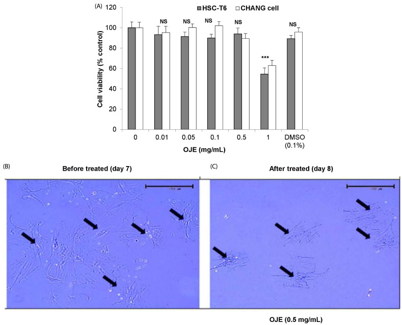
Fig. 1. Cell viability assay of HSC-T6/Chang liver cells and morphological changes in primary HSCs in response to treatment with OJE.
(A) HSC-T6 and Chang liver cells were incubated with OJE at the indicated concentrations for 24 h, after which the cell viability was determined by MTT assay. Primary HSCs were cultivated for 1 week (B) and exposed to OJE at 0.5 mg/mL for 24 h (C). Pictures were taken before and after 24 h of treatment with OJE. Magnification was 100×. Arrows indicate HSCs. The data are expressed as the means ± SEM (n = 8), which were compared using one-way analysis of variance (ANOVA) followed by Student's t-test. NS, not significant and ***P < 0.001 compared to the control group. OJE, O. japonica extract; HSC, hepatic stellate cells; DMSO, dimethyl sulfoxide.

