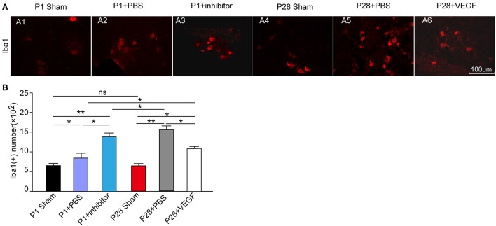
Figure 6.
Protective role of VEGF against inflammation in spinal cord in rats after ST. (A) Immunofluorescence staining for Ibal-1 to label microglia (red, A1–A6) in the L3 segment of the rat spinal cord; (B) The number of Ibal 1-positive cells increased in the four injured groups compared with that in the sham groups. There was no significant difference in the Ibal-1 positive cell number between the P1 and P28 sham groups. Among the four injured groups, the number in the P28 + PBS group was significantly higher than that in the P1 + PBS group or P28 + VEGF group, the number in the P1 + inhibitor group significantly increased compared with that in the P1 + PBS group. *P < 0.05; **P < 0.01; n = 6.

