


Figure 4.
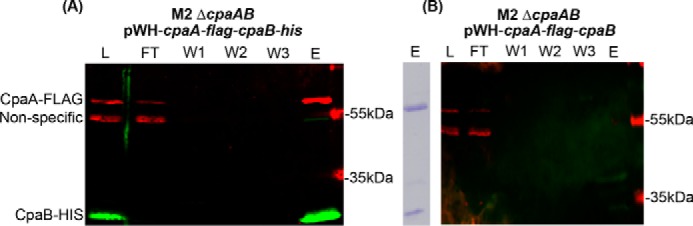
CpaA and CpaB strongly interact physically. A and B, Western blot analysis of nickel affinity purifications from cell lysates of M2 ΔcpaAB expressing either pWH-cpaA-flag-cpaB-his (A) or pWH-cpaA-flag-cpaB (B). Note that, in B, CpaB does not contain a C-terminal hexahistidine tag. CpaA-FLAG was detected using an anti-FLAG monoclonal antibody, whereas CpaB was detected using anti-histidine polyclonal antibodies. The right panel in A shows Coomassie-stained SDS-PAGE of the elution from A and thus represents the CpaA–FLAG–CpaB–His complex. L, loaded cell lysate, FT, flow-through, W1–3, washes one to three; E, elution.