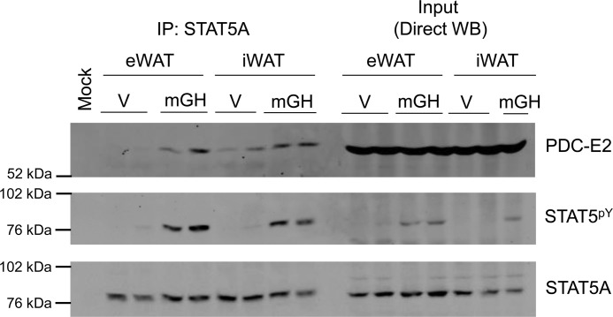
Figure 5.
STAT5A interacts with PDC-E2 in adipose tissue in vivo. Male C57BL/6J mice (12 weeks of age on a chow diet) were injected intraperitoneally with mGH (1.5 mg/kg) or vehicle (V: 0.1% BSA/PBS) 15 min prior to sacrifice. eWAT and iWAT white adipose tissue depots were collected in IP buffer and immediately placed on ice. eWAT and iWAT were homogenized using a Potter-Elvehjem tissue homogenizer and centrifuged. Floating lipid was removed, and the supernatants were used for IP and direct Western blotting experiments. IP experiments were performed using the anti-STAT5A antibody and 600 μg of tissue homogenate protein. The mock experiment contained IP antibody but no homogenate. Western blotting was used to examine the protein content of the immunoprecipitates (IP: STAT5A; left) and homogenate inputs (Direct WB; 150 μg of protein/lane; right). This experiment was performed one time, and two mice per condition were examined.

