
Figure 6.
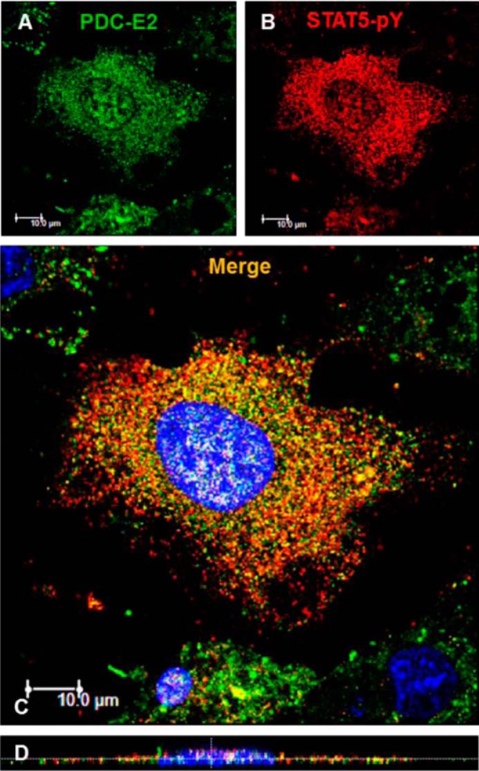
PDC-E2 interacts with STAT5pY in adipocyte nuclei. Mature 3T3-L1 adipocytes were seeded on coverslips, serum-deprived overnight, and then treated with 5 nm mGH for 20 min. After fixation, IF microscopy was conducted using primary antibodies directed against PDC-E2 (mouse primary) and STAT5pY (rabbit primary). A Dylight-488-conjugated anti-mouse secondary antibody (green; A) and an AlexaFluor 594-conjugated anti-rabbit secondary antibody (red; B) were used. Nuclei were counterstained with DAPI (4′,6-diamidino-2-phenylindole, a fluorescent stain that strongly binds DNA and stains the nucleus blue). Cell staining was imaged on a Leica TCS SP5 confocal laser-scanning fluorescence microscope using a ×63 oil immersion objective. The laser wavelengths were 405 nm (DAPI), 488 nm (green channel, 30% intensity; A), and 561 nm (red channel, 80% intensity; B). C, two-dimensional images from all three channels were merged. D, orthogonal sections were imaged using the z-stack feature (15 sections were scanned). This experiment was repeated at least two times on independent batches of cells.