
Figure 8.
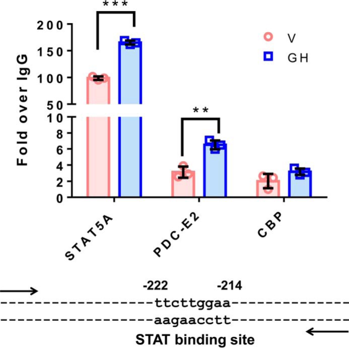
PDC-E2 binds to a STAT5 DNA-binding site within the cish promoter. Fully differentiated 3T3-L1 adipocytes were serum-deprived overnight and then treated with vehicle (V; NaHCO3) or 5 nm mGH for 20 min. Chromatin was cross-linked using DSG and 1% formaldehyde, and nuclear extracts were subjected to ChIP using anti-STAT5A, CBP, or PDC-E2 antibodies. The cish promoter DNA bound to each immunoprecipitated protein was quantified by qPCR, and percent input was calculated. Non-immune isotype-specific IgGs were used as negative controls, and all data were normalized against IgG binding and represented as fold over IgG. For each antibody, GH treatments (blue bars) were compared with vehicle (pink bars), and statistical significance was determined using the Student's t test and assigned as **, p < 0.005, and ***, p ≤ 0.001. These results have been replicated well over three times using independent batches of adipocytes. The cish gene is a well-known STAT5 target gene, and the STAT-binding site that we examined within the cish promoter is shown below the graph. The numbers above the binding site identify the location of the STAT-binding site relative to the cish transcription start site.