Fig. 1.
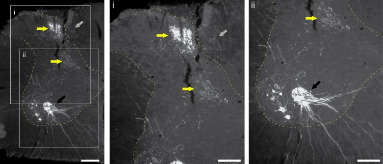
Histological example of phrenic afferent labeling in the C4 spinal cord. Cholera toxin β-subunit (CT-β) was applied to the phrenic nerve in an adult Sprague-Dawley rat using the method published by Nair et al. (2017). Tissues were harvest after 96 h and immunochemically processed to enable visualization of CT-β labeling. Phrenic motoneuron labeling is clearly observed (black arrows). A dense pocket of afferent projections is observed in the dorsal horn laminae II and III and projecting into the deeper lamina (yellow arrows). Afferent fibers are also labeled in the dorsal columns (gray arrows). Scale bars: 200 µm (left) and 50 µm (enlarged insets i and ii).

