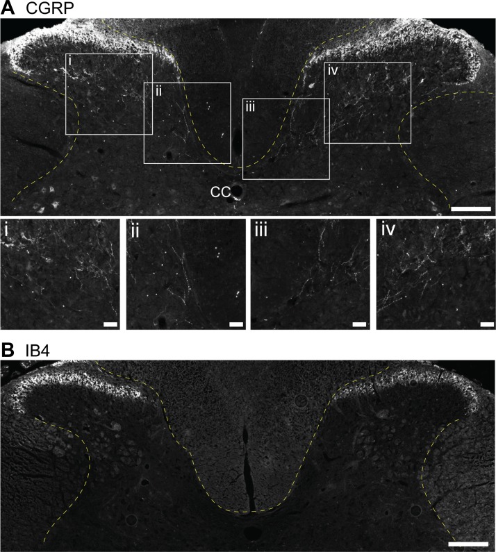
Fig. 2.
Histological example of calcitonin gene-related peptide (CGRP) and isolectin-B4 (IB4) immunochemical staining in the C4 spinal cord. Tissue sections from an adult Sprague-Dawley rat were immunochemically stained using the method published by Nair et al. (2017). A: CGRP staining is prominent in dorsal horn laminae I and II, and CGRP-positive fibers can be seen extending to deeper laminae. B: IB4 immunoreactivity is concentrated in lamina II but is absent from the deeper laminae. Scale bars: 200 µm (A and B) and 50 µm (enlarged insets i–iv).

