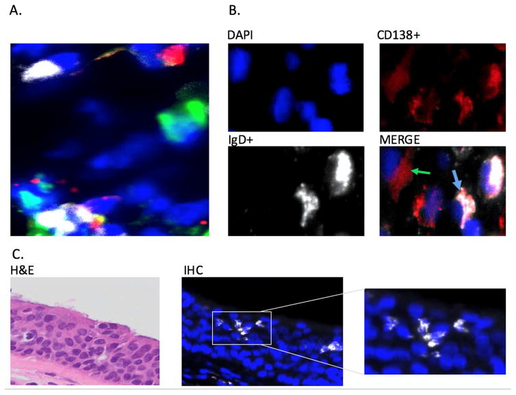
Figure 1.
A. Immunofluorescent triple staining for cells expressing cytoplasmic IgD (white), IgM (red), and IgA (green) in sinus tissue. Nuclei are stained with DAPI (blue). Results are shown from a representative patient with CRSsNP. Original magnification: 400x. B. Immunoflourescent staining showing co-expression of cytoplasmic IgD (white), plasma cell marker CD 138 (red) and nuclear staining (blue). Blue Arrow: IgD/CD138 Costaining. Green Arrow: CD138 without IgD staining. Results shown are representative of 18 samples. Original magnification: 400x. C. Localization of IgD plasma cells in the sinus epithelium. IgD+ plasma cells: White, DAPI nuclear stain: Blue. Inset: higher magnification showing morphology of IgD+ plasma cells Original magnification: 400x.

