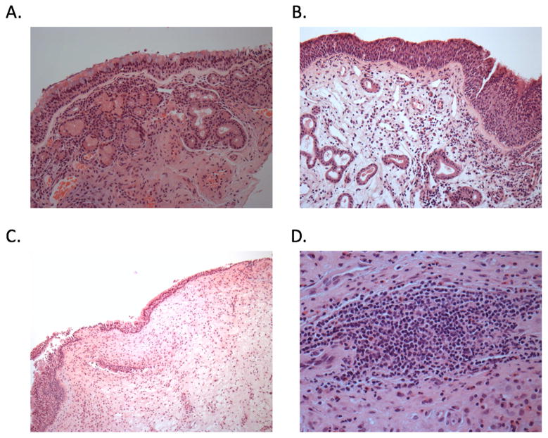
Figure 4.
H&E staining of sinus tissue. A. Normal control: Normal sinonasal mucosa lined by ciliated pseudotratified, columnar epithelium with identifiable mucocytes (goblet cells) with underlying seromucous glands scattered throughout the submucosa. Magnification 200x B. CRSsNP: Submucosal edema with mixed inflammatory cells, including mature lymphocytes with variable plasma cells, eosinophils, histiocytes, and neutrophils. Magnification 200x. C. CRSwNP: The surface epithelium is composed of intact respiratory epithelium. The underlying stroma is markedly edematous and is noteworthy for the absence of seromucous glands. A mixed chronic inflammatory infiltrate is present and is predominantly composed of eosinophils, plasma cells, and lymphocytes. Magnification 100x D. Lymphoid follicle: Dense and sizable (>50 lymphoid cells) aggregate with predominantly lymphoid cells (i.e. other chronic inflammatory including plasma cells, eosinophils, histiocytes, and neutrophils cells constitute only a minor subset within the aggregate). All other lymphoid patterns are considered a part of the chronic inflammatory infiltrate. Magnification 400x.

