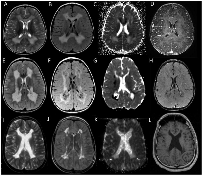
Figure 2. Evolution of lesions in SDH-related leukoencephalopathy.

(A-D) Top Row – Early stage MRI. The abnormal white matter looks swollen, especially the corpus callosum. Myelin micro-vacuolization is suspected in areas of restricted diffusion seen on ADC maps. There is no enhancement. (E-H) Middle Row – Intermediate stage MRI. Tissue necrosis is suspected based on MRI findings with white matter rarefaction seen on FLAIR imaging and small foci of contrast enhancement. Areas of low ADC values continue to be present. (I-L) Bottom Row – Late stage MRI. Atrophy and collapse of the affected white matter is seen, with cysts. ADC values are low and no further contrast enhancement is present. (A,E,I) T2-weighted imaging from select patients. (B,F,J) FLAIR imaging from select patients. (C,G,K) Diffusion weighted imaging from select patients. (D,H,L) Contrast enhanced images from select patients.
