Figure 1.
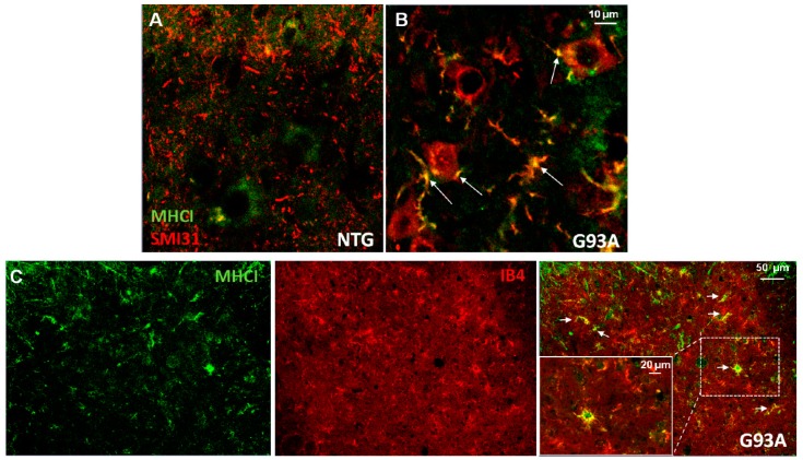
Major histocompatibility complex class I (MHCI) immunoreactivity in dystrophic neurites of motoneurons (MNs) and activated microglia. (A,B) Representative immunofluorescent microphotographs showing MHCI (green) and phosphorylated neurofilaments (SMI31) in L3–L4 ventral horn of normal control and SOD1G93A mice at the early phase of the disease. Note a diffuse cytosolic MHCI immunostaining in MNs of wild-type mice, while SMI31 appears only as punctiform staining of axons of different calibers in the spinal cord parenchyma (A). In SOD1G93A mice, SMI31 immunolabeling is prevalent in MN somata and in dystrophic neurites/axons around MNs and partially co-localizes with MHCI (white arrows—yellow). (C) Representative immunofluorescent microphotographs showing the colocalization of MHCI (green) and activated microglia (Ib4) in the ventral horn of SOD1G93A mice at the early phase of the disease (white arrows—yellow). Note in the magnification the presence of MHCI in reactive microglia.

