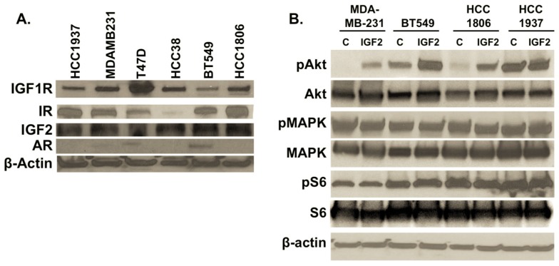
Figure 2.
(A) Expression of IGF2, IGF1R, insulin receptor (IR) and androgen receptor (AR) in TNBC cultures. Total protein was isolated from cell cultures. Forty micrograms of protein were separated and transferred to PVDF membranes for detection of IGF1R (1:500, Cell Signaling #3027, Danvers, MA, USA), IR (1:500, Cell Signaling #3025), IGF2 (1:1000, AbCam ab9574), and AR (1:500, Cell Signaling #5153). β-actin (1:2000, Sigma #A1978, St. Louis, MO, USA) was used as a loading control. TNBC cells include HCC1937, MDA-MB-231, HCC38, BT549 and HCC1806, with ERα-/PR-positive T47D cell line as a control; (B) Effects of IGF2 treatment on downstream phosphorylation of MAPK, AKT and S6. IGF2-induced activation of IGF1R leads to increased phosphorylation of AKT in most TNBC cells assessed. TNBC cultures were treated with IGF2 (100 ng/mL) in serum- and phenol red-free media for 20 min. Total protein was isolated, separated and transferred to PVDF membranes. Detection of MAPK (1:1000; Cell Signaling #9102), pMAPK (Cell Signaling #4370), S6 (1:2000; Cell Signaling #2217), pS6 (Cell Signaling #4858), AKT (1:1000 Cell Signaling, #4685) and pAKT (Cell Signaling #4060) was accomplished following the manufacturer’s recommended protocols (Methods). C = control vehicle-treated cells. IGF2 = cells treated with IGF2 for 20 min. Western immunoblots are representative of three independent experiments.

