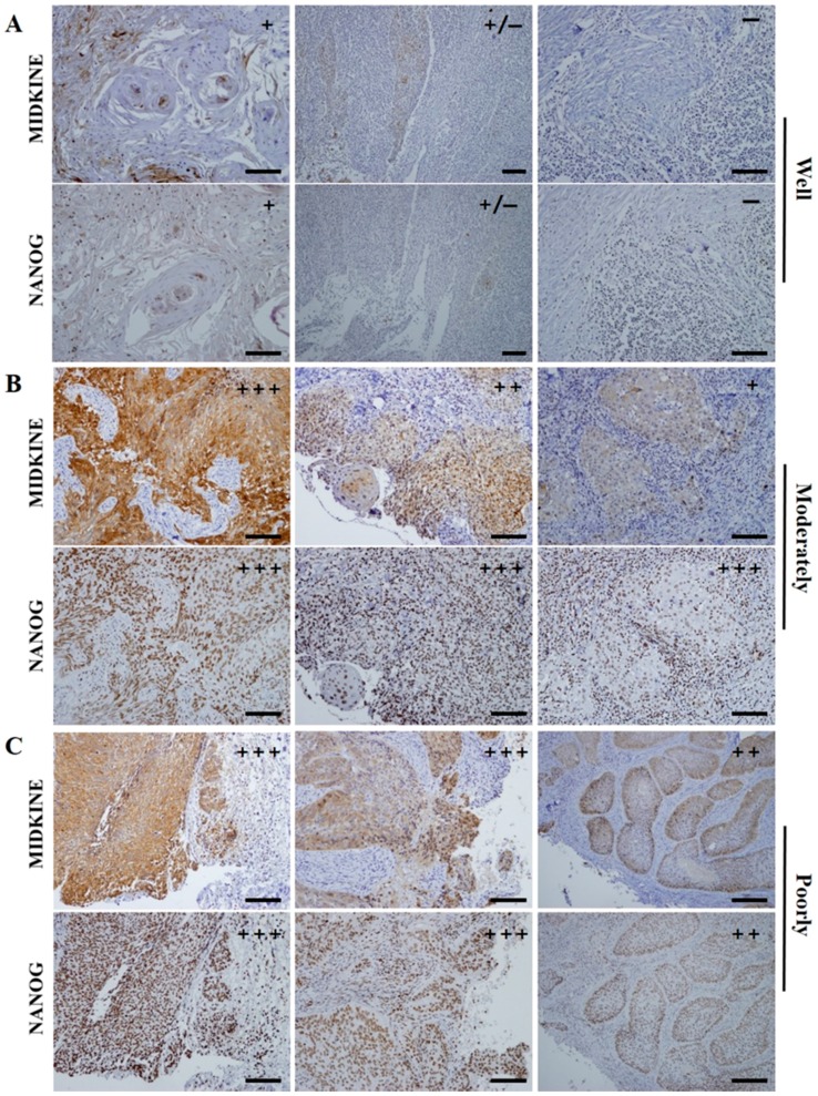
Figure 2.
Representative serial section images of IHC staining of MK and NANOG in the pretreatment OSCC biopsy specimens as their histopathological grades. (A) In well differentiated OSCCs, weak or negative expression of MK and NANOG is usually detected; (B,C) high-grade tumors, moderately- or poorly-differentiated OSCCs, dominantly revealed the enhanced (strong and moderate) expressions of MK and NANOG proteins. The expression patterns these two marker proteins, including immunostaining intensity and positive cell ratio, revealed highly similar features in OSCC specimens. In particular, enhanced co-localization of MK and NANOG proteins was usually detected in the cancer cells of high-grade tumors. Scale bar = 100 µm.

