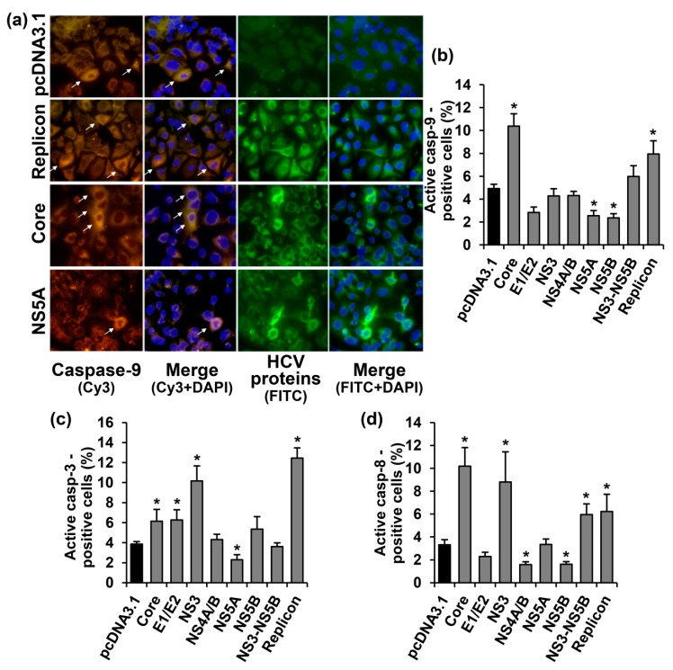
Figure 2.
HCV proteins affect activation of caspases-3, -8 and -9 in Huh7.5 cells in different manners. (a) Immunofluorescent staining of the activated caspase-9 and HCV proteins in Huh7.5 cells transiently expressing the HCV core or NS5A proteins, or harboring the full-length HCV replicon (400× magnification). Vertical panels left to right: staining with rabbit anti-caspase-9 primary and anti-rabbit secondary antibodies conjugated to Cy3 (orange), merge with nuclear staining with DAPI (blue), staining with mouse monoclonal antibodies to HCV proteins and anti-mouse secondary antibodies conjugated to fluoresceine isothiocianate (FITC; green), combined with nuclear staining with DAPI (blue). The arrows indicate caspase-9 positive cells. (b–d) Percentages of the cells which tested positive for the caspases-9 (b), -3 (c), and -8 (d). Values on each diagram are means ± SEM of eight measurements done in three independent experiments, * p < 0.05 compared to the cells transfected with the empty vector (black bar).

