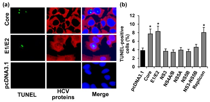
Figure 3.
The HCV core and E1/E2 increase the number of Huh7.5 cells with nuclear DNA fragmentation, i.e., at the end stage of apoptosis. (a) Huh7.5 cells transfected with the core- and E1/E2-expressing plasmid or the empty pcDNA3.1 vector were stained 72 h posttransfection with the “DeadEnd™ Fluorometric terminal deoxynucleotidyl transferase dUTP nick end labeling (TUNEL) System” kit (green), with mouse monoclonal antibodies on HCV proteins and anti-mouse secondary antibodies conjugated to Alexa Fluor 594 (AF594), and with DAPI. Vertical panels left to right: TUNEL staining (green), HCV proteins (red), and overlay of TUNEL, HCV proteins and DAPI staining; (b) Percentages of TUNEL-positive cells. Values are means ± SEM of eight measurements done in three independent experiments, * p < 0.05 compared to the cells transfected with the empty vector (black bar).

