Figure 4.
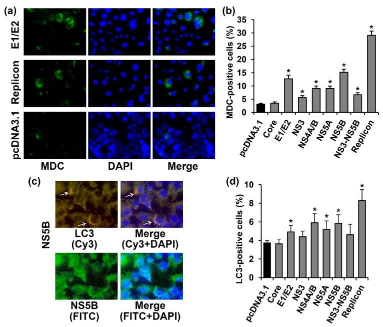
HCV proteins activated autophagy in Huh7.5 cells, as revealed by the enhanced incorporation of monodansylcadaverine into autophagosomes and detection of LC3. (a) Huh7.5 cells harboring the HCV replicon, or transfected with the E1/E2-expressing plasmid or the empty pcDNA3.1(+) vector were stained 72 h posttransfection with the monodansylcadaverine (MDC) and with DAPI. Vertical panels from the left to the right are: MDC staining (green), nuclear staining with DAPI (blue), overlay of MDC and DAPI staining; (b) Percentages of MDC-positive cells. (c) immunofluorescent staining of the activated LC3 and NS5B protein in Huh7.5 cells (400× magnification). Vertical panels left to right: staining with rabbit anti-LC3 primary and anti-rabbit secondary antibodies conjugated to Cy3 (orange) or primary mouse anti-NS5B antibody and secondary anti-mouse antibodies conjugated to FITC (green), merge with nuclear staining with DAPI (blue) The arrows indicate cells with LC3 punctuate staining; (d) Percentages of LC3-positive cells. Values are means ± SEM of eight measurements done in three independent experiments, * p < 0.05 compared to the cells transfected with the empty vector (black bar).

