Figure 2.
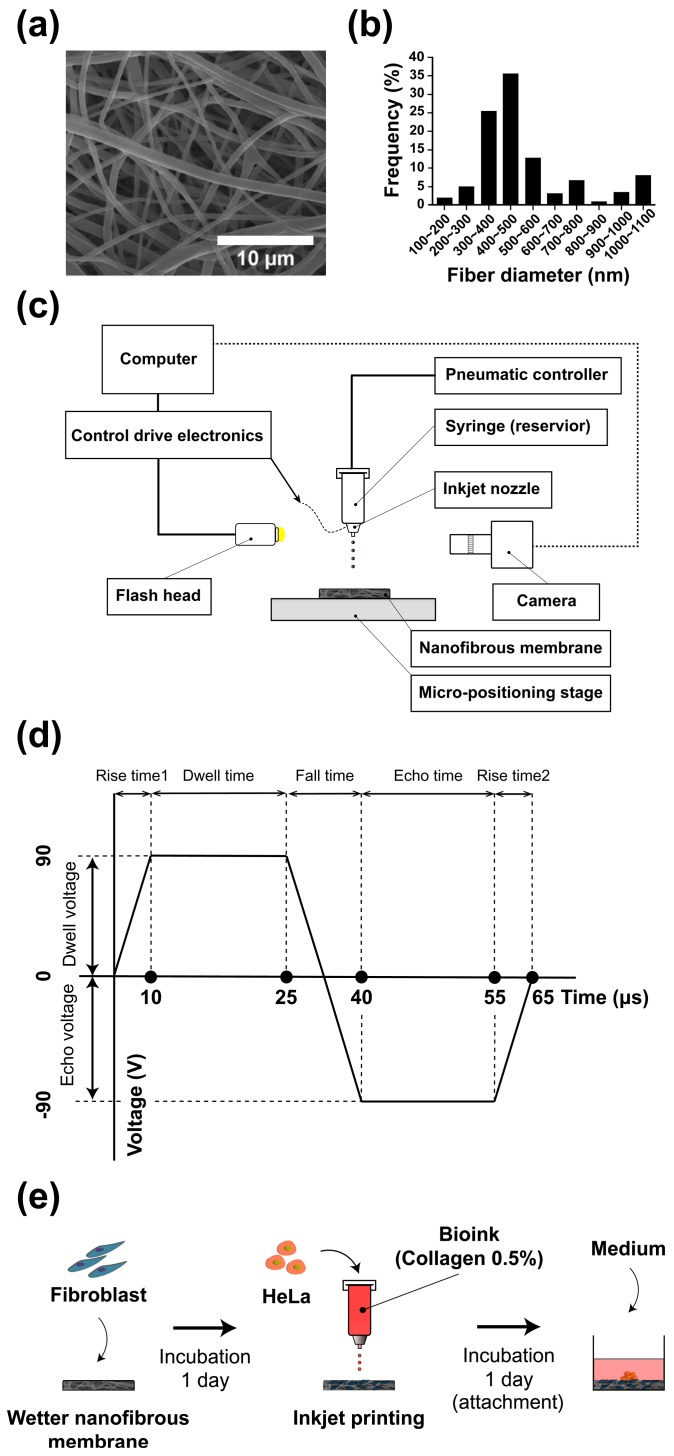
Scanning electron microscopy (SEM) image of the electrospun nanofibrous membrane (NF) (a); diameter distribution of the nanofibers (b); schematic representation of the inkjet printer (c); bipolar waveform for piezo actuation (d); and schematic representation of the cancer microtissue fabrication process (e).

