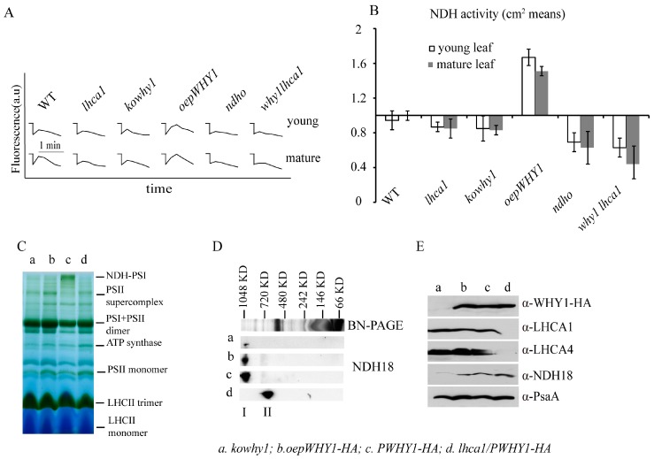
Figure 5.
The stability of NDH-PSI complexes of thylakoid membranes and NDH activities in the different WHY1 and lhca1 mutants. (A) Determination of NDH activity by chlorophyll fluorescence analysis. Five-week-old plants including different WHY1 and lhca1 mutants, why1lhca1 double mutant and wild-type (WT) were illuminated for 5 min with AL (200 μmol m−2 s−1). After illumination, the subsequent transient increase in chlorophyll fluorescence were recorded as an indicator of NDH activity (a.u.; arbitrary units). The fluorescence levels were standardized by the Fm level. Curves shows trace of chlorophyll fluorescence in mutants and WT plants. ndho line was used as positive control; (B) Quantification of NDH activity measured in (A). The transient increase in chlorophyll fluorescence in mutants was quantified by comparing the peak area with that of WT. The peak area of WT was set as 1. Data presented are the mean of ± SE (n = 5–6); (C) Blue Native PAGE of thylakoid membrane, Band I is the NDH-PSI supercomplex detected in PWHY-HA line; (D) 2D SDS-PAGE separation and immunodetection of NDH-PSI supercomplex containing NDH18 protein (I) and monomer NDH18 (II); (E) Immunodetection of thylakoid membrane, the antibody against to HA peptide, LHCA1, LHCA4, NDH18 (provided by Shikanai group, Japan) and PsaA in kowhy1 (a), oepWHY1-HA (b), PWHY1-HA (c), and PWHY1-HA in lhca1 background, lhca1/PWHY1-HA (d).

