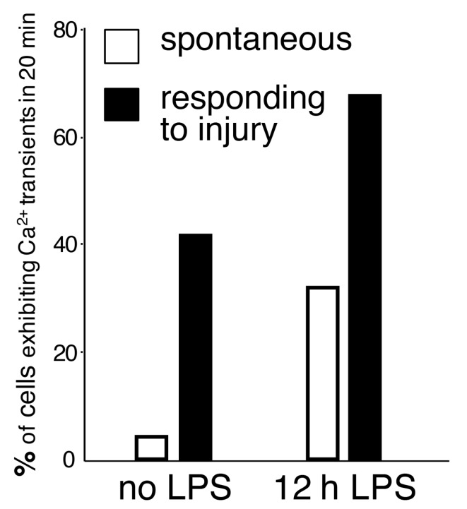
Figure 2.
The frequency of spontaneous and evoked Ca2+ transients in cortical microglia, calculated as a percentage of cells exhibiting at least one Ca2+ spike during a 20-min recording session. Ca2+ activity was detected with GCaMP5G and two-photon laser scanning microscopy. In this model, only 4% of resting microglia exhibited spontaneous Ca2+ activity (no LPS, white bar). The frequency of Ca2+ transients increased 8-fold after LPS exposure (12 h LPS, white bar). In cells extending processes towards the focal laser injury, 67% of microglia displayed Ca2+ activity (12 h LPS, black bar). Adapted from [16].

