Figure 1.
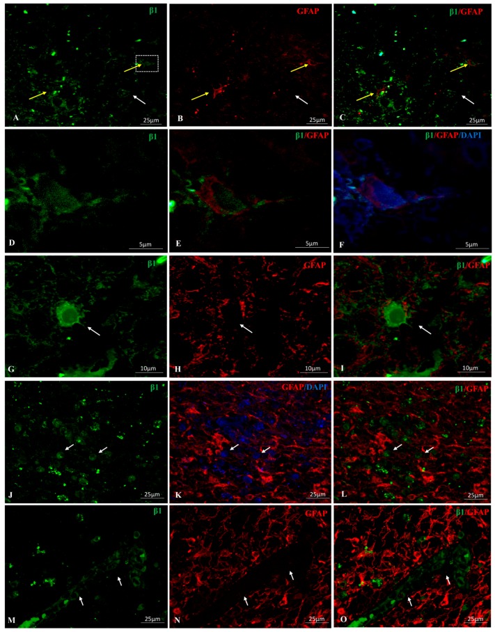
Double immunolocalization for GFAP (Glial Fibrillary Acidic Protein) (red) and Na-K-ATPase β1 subunit isoform (green) in primary (A–I) and secondary (J–L) glioblastoma multiforme (GBM). (A–C): Yellow arrows point to β1/GFAP positive cells. Faint β1 positive staining is observed in the nucleus of the cell located in the right side of the image (enlarged in panels (D–F)). White arrow points to a GFAP-cell expressing β1 in plasma membrane, nucleus and podosome-like structures. (G–I): β1+ immunostaining in cytoplasm, membrane and nuclear envelope of a giant cell. Arrow points to an invadosome β1+. The cell is filled by GFAP+ filaments. (J–O) Secondary GBM. β1 signal in the nuclear envelope and, sometimes, nucleosol of GFAP− cells (arrows). Note the brighter fluorescence signal for GFAP in secondary over primary GBM. (M–O) Arrows point β1+ stromal and microenvironment cells, GFAP−.

