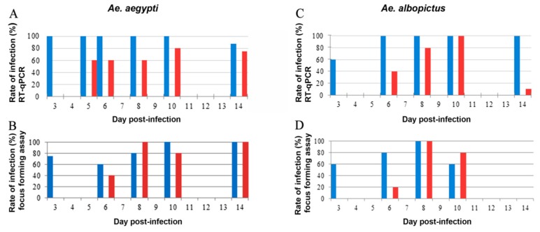
Figure 1.
Rate of infection detected by RT-qPCR and immunochemical plaque assay in midguts (MG) and salivary glands (SG) of Ae. aegypti and Ae. albopictus. MG (blue bars) and SG (red bars) were analyzed by RT-qPCR (A,C) and titration (B,D) from Ae. aegypti (A,B) and Ae. albopictus (C,D). Ae. aegypti MG and SG were sampled on days 3, 5, 6, 8, 10, and 14, whereas Ae. albopictus MG and SG were sampled on days 3, 6, 8, 10, and 14. The graphs present the results of one experiment for which 5 mosquitoes were tested for each time point. It was repeated two times.

