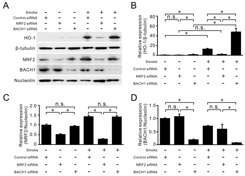
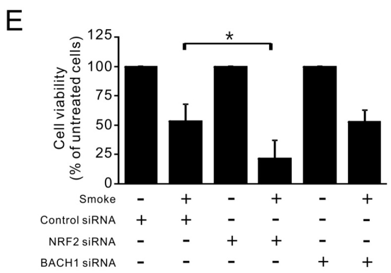
Figure 5.
Contributions of NRF2 and BACH1 to HO-1 protein level and cell viability in response to smoke exposure. (A) HBE1 cells transfected with siRNA targeting NRF2 and BACH1 were subsequently exposed to cigarette smoke. After 24 h of exposure, cells were collected for protein analysis by immunoblotting. (B–D) The relative expression of HO-1/β-tubulin (B); NRF2/Nucleolin (C); and BACH1/Nucleolin (D) were quantified from three separate experiments as fold relative to that of untreated HBE1 cells with control siRNA. * p < 0.05 (n = 3; mean ± S.D.); (E) HBE1 cells with NRF2 or BACH1 siRNA transfection were exposed to cigarette smoke for 72 h and cell viability was determined by MTS assay (n = 3; mean ± S.D.). * p < 0.05.


