Figure 3.
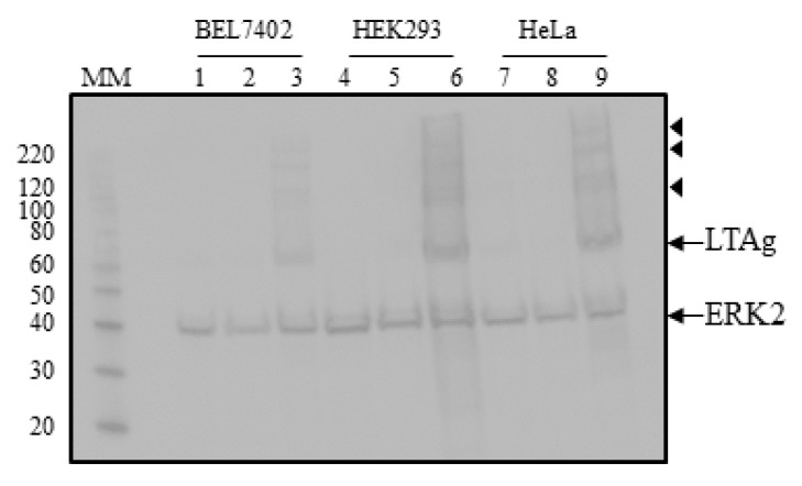
Detection of ectopic expressed HPyV9 LTAg in BEL7402, HEK293, and HeLa cells. Lanes 1–3: lysates of BEL7402 cells, lanes 4–6: lysates of HEK293 cells; lanes 7–9: lysates of HeLa cells. Lanes 1, 4, and 7: mock transfected cells; lanes 2, 5, and 8: cells transfected with empty vector pcDNA3.1(+); lanes 3, 6, and 9: cells transfected with expression plasmid for hemagglutinin (HA) tagged LTAg of HPyV9. Western blots were performed with anti-HA and anti-ERK2 antibodies. The molecular mass (in kDa) of the markers (lane MM) is indicated. The arrows indicate the bands corresponding to HPyV9, LTAg, and ERK2, respectively, while the bands marked with arrowheads may represent post-translationally modified HPyV9 LTAg.

