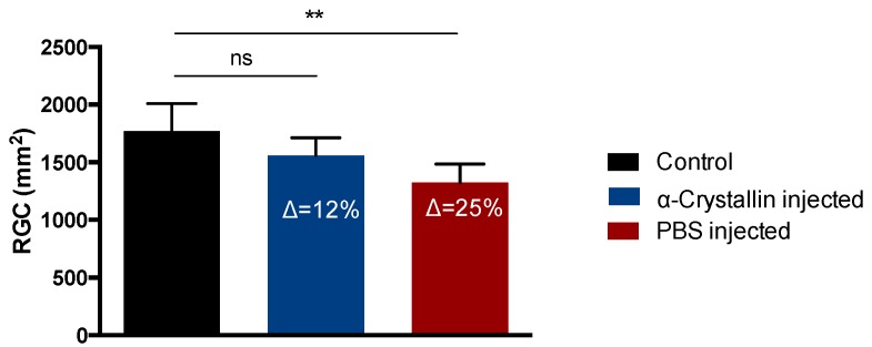
Figure 2.
Quantification of RGC density in retinal flat-mounts. Elevation of the IOP resulted in an average loss of ∆ = 12% retinal ganglion cell (RGCs) in the α-crystallin B injected animals compared to the untreated contralateral eyes, while the RGC loss in the PBS-injected animals was about 25% (** p < 0.01, ns—not significant, n = 11, mean ± SD, one-way ANOVA).

