Figure 1.
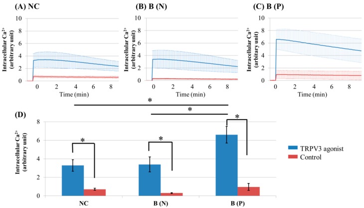
Ca2+ influx in cultured keratinocytes of: (A) Normal control; (B) Nonpruritic burn scar and (C) pruritic burn scar; (D) Intracellular Ca2+ levels at the time of TRPV3 agonist treatment. Each group was treated with TRPV3 agonist (500 μM Carvacrol and 200 μM 2-APB mixture) and Ca2+ buffer with 0.5 M ethylenediaminetetraacetic acid (EDTA) was used for control. The Ca2+ influx in cultured keratinocytes decreased slowly with time. NC: keratinocytes from normal control; B (N): keratinocytes from a nonpruritic burn scar; B (P): keratinocytes from a pruritic burn scar; Error bars in (A–C): standard deviation of the mean value obtained from three experiments; Error bars in (D): standard error, each performed in triplicate.* p < 0.001.

