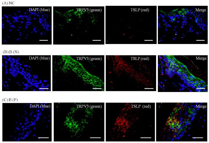
Figure 9.
Three-color immunofluorescence confocal images were obtained for DAPI (blue), TRPV3 (green) and TSLP (red) from tissues of (A) normal, (B) nonpruritic and (C) pruritic burn scars. The staining of TRPV3 and TSLP, well observed in spinous layer of the epidermis, was least in (A) normal control and greater in (B,C) burn-scar tissues with or without pruritus. NC: keratinocytes from normal control; B (N): keratinocytes from nonpruritic burn scar; B (P): keratinocytes from pruritic burn scar; TSLP: thymic stromal lymphopoietin. Scale bars = 50 μm.

