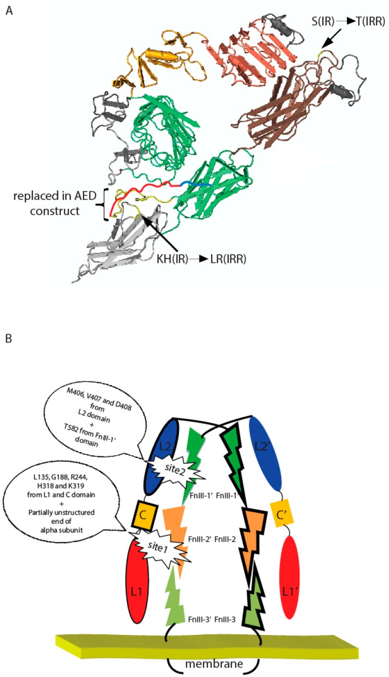
Figure 5.
(A) Positions of indicated T582, L868, R869, and region (646–716) within fibronectin repeats domains and their IR substitutes on a putative three-dimensional model of IRR built according to the described structure of IR ectodomain [23] with Cn3D 4.3.1 software (NCBI, Bethesda, MD, USA). Region, which were swapped in AED chimera construct, was indicated by yellow color (presented in IR ectodomain structure) and red color (unstructured part of IR alpha subunit). Blue color showed rest of unstructured part of IR alpha subunit. (B) Two-site activation model of IRR. Important residues are indicated. Domains indicated by different colors. Monomers indicated by trait.

