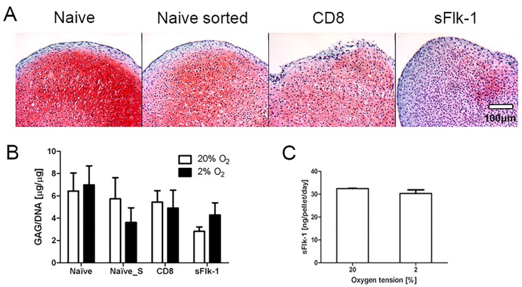
Figure 2.
In vitro chondrogenesis of nasal chondrocytes. (A) Representative Safranin-O staining images of NC cultured in pellets at 20% of oxygen tension. Naïve, naïve mock-sorted (Naïve sorted), CD8 (control), and sFlk-1-releasing NC were cultured in vitro in pellets and analyzed for their chondrogenic potential. (B) Glycosaminoglycan (GAG) content of pellets generated by NC cultured at either 2% or 20% of oxygen tension (2 donors, n = 9). (C) Amount of mouse sFlk-1 released by sFlk-1-expressing NC-based pellets cultured at different oxygen tensions (2 donors, n = 4). No statistical significant difference has been found.

