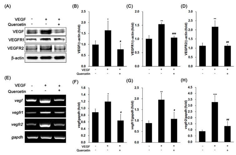
Figure 2.
Effect of quercetin on the expression of vascular epithelial growth factor (VEGF), VEGFR1, and VEGFR2 in VEGF-stimulated 661W cells. (A) Cells were treated with VEGF (20 ng/mL) in the absence or presence of quercetin (0.1 µM) for 16 h. The protein expression was detected by immunoblot. (B–D) Densitometry quantifications of VEGF (B), VEGFR1 (C), and VEGFR2 (D) protein expression were measured by ImageJ. Data are the means ± SDs of three independent experiments. * p < 0.05 and ** p < 0.01 indicate significant differences compared to the non-treated control group. # p < 0.05, ## p < 0.01 and ### p < 0.001 indicate significant differences compared to the VEGF-only treated group. (E) Cells were treated with VEGF (20 ng/mL) in the absence or presence of quercetin (0.1 µM) for 6 h. Gene expression was determined by RT-PCR. (F–H) Densitometry quantifications of vegf (F), vegfr1 (G), and vegfr2 (H) gene expression were measured by ImageJ. Data are the means ± SDs of three independent experiments. * p < 0.05, ** p < 0.01, and *** p < 0.001 indicate significant differences compared to the non-treated control group. # p < 0.05 and ## p < 0.01 indicate significant differences compared to the VEGF-only treated group.

