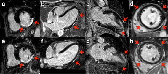
Fig. 3.

Selected matched images of 2D (upper row) and iNAV-3D (lower row) LGE in a patient with ischemic heart disease (a and e), myocarditis (b and f), hypertrophic cardiomyopathy (c and g) and dilated cardiomyopathy (d and h). Red arrows and star indicate the presence of LGE. Blurring due to residual respiratory motion is noticed in the latter three 3D–LGE images (f-h). Abbreviations as above
