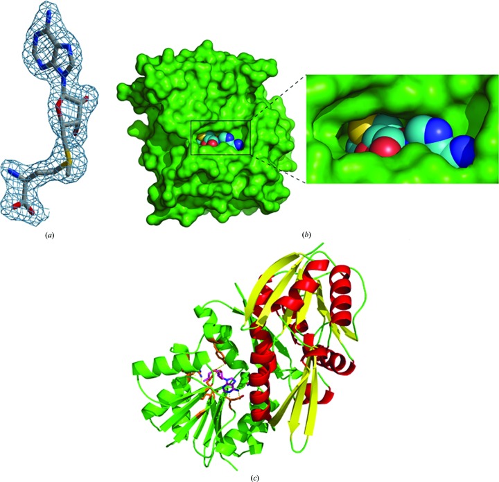
Figure 2.
(a) SAM (stick model) shown within a difference density map contoured at 1.3σ. (b) The binding pocket of the enzyme (green surface) with the SAM molecule shown as a sphere model. (c) The interaction of SAM (magenta stick model within the green monomer) with the other monomer (red α-helices and yellow β-sheets) leading to dimerization.

