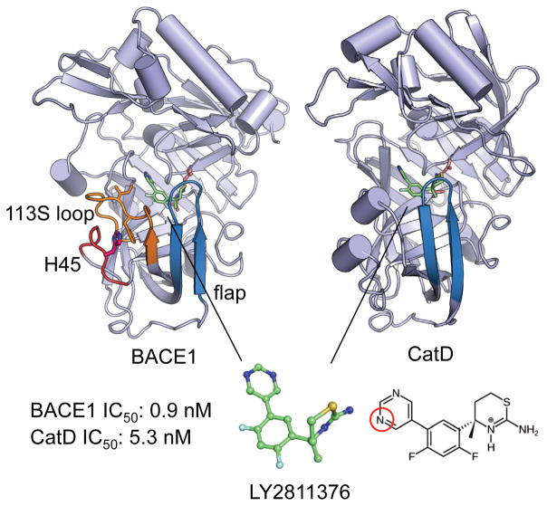
Figure 1. Structures of BACE1 and CatD bound to the inhibitor LY2811376.
In both structures, the catalytic dyad and inhibitor are drawn as sticks, while the flap is colored blue. In BACE1, the 113S loop18 and the loop that contains His45 are colored orange and red, respectively. The sidechains of His45, Phe109 and Ile110 are shown. In the inhibitor, nitrogen, fluorine and sulfur atoms are shown in blue, cyan and yellow, respectively. The endocyclic nitrogen on the aminothiazine ring carried a +1 charge, while a pyrimidine nitrogen (circled in red) was titratable in the simulation pH range. The coordinates for the BACE1 complex were taken from the X-ray crystal structure with PDB ID 4YBI;2 the coordinates for the CatD complex were taken from our previous work. 12 The listed IC50 values (at pH 4.6) were obtained by Pfizer3 and correspond to a selectivity of 6 fold or a binding free energy difference of 1.0 kcal/mol.

